


Varicose veins are an abnormal dilation of veins located on the surface, characterized by an increase in their diameter and length, which in turn leads to cylindrical, serpentine, saccular, and mixed-type lesions in the venous trunks. Today, varicose veins are a widespread pathology and women get sick more often than men, almost three times. This is mainly due to the anatomical features of the body and some of the load on the lower extremities during pregnancy.
As a general rule, varicose veins are primary and secondary. In the first variant, the disease is caused by an initial weakness of the wall of the great vein, which is localized under the skin, or by congenital dysfunction of the valves. The development of secondary venous pathology is influenced by deep vein thrombosis or acquired valve insufficiency due to pregnancy, high physical exertion, prolonged standing, etc.
As the hydrostatic pressure in the veins increases, the diameter of these vessels expands and the impaired functions of the valves worsen. All of this disrupts the blood circulation in the superficial veins, and due to the inadequate functioning of the peripheral veins, blood flows back from the deep veins to the saphenous veins, which become too elongated, writhing, forming various forms of expansion. In the future, due to the pronounced stagnation, the trophism of the tissues will be disturbed, and ulcers, eczema and dermatitis will develop.
The lower extremities are varicose veins
This disease is characterized by the formation of venous walls, sacral dilatation, serpentine twisting, increased length of the valves, and insufficiency.
As a general rule, varicose veins in the lower extremities occur in 20% of the population. In addition, it affects both boys and girls before puberty. But adult women are much more likely to be affected by varicose veins, unlike men. In addition, the number of patients increases with age. This can be explained by the rearrangement of the hormonal background of the female body during pregnancy and menstruation, which causes a weakening and dilation of the tone of the veins, some insufficiency of the valves of the communicating and saphenous veins, and the opening of the veins. arteriovenous shunts and circulatory disorders in the veins.
To date, the true cause of varicose veins in the lower extremities is not yet known. It is hypothesized that valve malfunction and increased venous pressure are associated with the etiological cause of the disease. Considering all the factors that predispose to the pathological process in the veins of the lower extremities, there are two types of varicose veins: primary and secondary.
Primary varicose veins on the surface are characterized by the presence of normal, deep veins. In the case of secondary varicose veins, various complications of the deep veins, arteriovenous fistulas, congenital absence or underdevelopment of venous valves play an important role.
Risk factors involved in the development of the lower extremity varicose veins: increased hydrostatic pressure in the trunk of the veins, thinning of the wall, impaired metabolic processes in the smooth muscle cells, movement of blood from the deep veins to the superficial ones. This inverse movement of blood in the form of vertical and horizontal reflux causes nodular dilation, elongation, and curvature of the superficial veins localized under the skin. The final link in the pathogenesis is the trophic venous ulcer of cellulitis, dermatitis, and leg.
The symptomatic picture of lower extremity varicose veins consists of complaints from patients about pre-existing varicose veins that cause cosmetic discomfort, some severity, sometimes lower extremity pain, nocturnal cramps, and trophic changes in the legs.
The extent of venous vessels can range from smaller "stars", from reticular nodules to coarsely writhing strains, and from nodes, plexuses, which are clearly visible in the vertical position of patients. Almost 80% is the lesion of the trunk and branches of the great vein on the surface, and 10% is the saphenous vein of the small vein. In addition, in 9% of patients, lesions of both veins were involved in the pathological process.
As a result of a progressive process, the patient begins to experience rapid fatigue, some severity and tension in the legs, cramps in the calf muscles, swelling of the legs and feet, and paraesthesia. In addition, the legs usually swell in the late afternoon, but after sleeping, this swelling goes away.
Varicose veins are often complicated by acute thrombophlebitis of the veins on the surface, with redness, cord-like, painful vein compression characterized by dilation, and periphlebitis. Very often, the varicose veins rupture as a result of minor injuries, and this leads to bleeding. As a general rule, blood from a cracked lump can flow in streams and the patient sometimes loses quite a large amount of it.
In addition, there is no difficulty in diagnosing lower extremity varicose vein and joining the CVI based on patient complaints, a history of the disease, and objective test results.
An essential value in making a diagnosis is the ability to determine the condition of the valves of the main and communicative veins and to assess the permeability of the deep veins.
Causes of varicose veins
This pathological process is characterized by dilation of the veins on the surface of the subcutaneous tissue and is accompanied by insufficient work of the valves in the veins and damage to the blood circulation in them. Varicose veins are one of the most common vascular diseases in the working age population.
There are usually a number of predisposing factors in the development of the disease as well as its progression. The definite contribution of inheritance to the appearance of varicose veins has not yet been proven. The development of this pathological process can currently be influenced by conditions caused by changes in diet, lifestyle and hormonal background.
In addition, the occurrence of this pathological process is related to the improper organization of the work process. Many, depending on their work, spend a significant amount of time standing or sitting, and this has a rather bad effect on the valve apparatus of the veins in the lower extremities. In addition, work involving heavy physical work is considered unfavorable, especially in the form of a jerky load on the legs during weight lifting.
Nowadays, long-distance travel or flights that contribute to venous congestion of the blood in the legs and risk factors for the development of venous pathologies negatively affect the blood flow system in the veins. In addition, wearing tight underwear causes the lumbar veins to compress and the corset increases the pressure inside the peritoneum, so it is not recommended to wear it constantly. This also applies to high heels in the presence of uncomfortable footrests.
Repeated pregnancy has been shown to be a risk factor for varicose veins. This can be explained by the fact that an enlarged uterus increases the pressure inside the peritoneum and progesterone destroys the elastic and collagenous fibers in the vein wall. In addition, diseases such as rheumatoid arthritis, osteoporosis, and changes in hormonal status increase the risk of developing this pathological process.
Typical causes of varicose veins are peculiarities of their structure on the lower extremities. On the surface is a system of veins, i. e. , the saphenous veins, such as the system of small and large veins, and the deep veins of the thighs and legs, and the perforating veins connecting the two previous systems. With normal blood circulation, blood flow to the lower extremities occurs in 90% of the deep veins and 10% of the superficial veins. But in order for the blood to move toward the heart, and not the other way around, there are valves in the venous walls that strike and prevent the blood from flowing from top to bottom under the force of gravity. Muscle contractions are also of great importance, contributing to normal blood flow. In addition, in a vertical position, blood stagnation develops, and the pressure in the veins begins to increase, leading to dilation. In the future, the valves will not work properly, which will be the reason for the valve plates not closing due to the incorrect movement of blood from the heart.
The keys of deep veins were touched particularly quickly due to the maximum load applied to them. And with a system of perforating veins to reduce overpressure, blood flows into veins under the skin that are not formed in large quantities. All this leads to the overhang of the venous walls, resulting in the formation of characteristic varicose nodes. However, the increased amount of blood continues to flow into the deep veins, resulting in insufficiency in the valves of the perforating veins without some obstruction of blood flow in a horizontal position, first to the deep vessels and then to the superficial ones. And finally, CVI develops with manifestations such as edema, pain, and trophic ulcers.
Symptoms of varicose veins
Varicose veins are characterized by the expansion of veins localized under the skin in the form of saccharoid or cylindrical lesions. With this pathological disease, veins twisting appear on the surface of the skin of the legs and feet. The maximal appearance of varicose veins occurs after long or strenuous physical exertion. In young women, it is quite common for veins to dilate during or after pregnancy.
The early stages of varicose veins are characterized by few and non-specific symptoms. At this point, patients get tired quickly, with constant difficulty in their legs, burning, cracking, especially after physical exertion. In addition, transient edema and painful pains sometimes appear along the entire length of the veins. However, after prolonged static loading in the late afternoon, the back of the ankle and leg swells. Some features of edema are that they disappear in the morning after a night’s rest. There are usually no visible signs of varicose veins at this stage. However, these early-stage symptoms should serve as an indication to the patient to consult a specialist to prevent progression of varicose veins.
This disease is characterized by slow, sometimes decades of development. Therefore, the development of varicose veins results in CVI (chronic venous insufficiency) as a result of poor treatment.
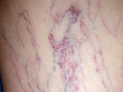
An important symptom of the disease is also the spider veins, which are a spider web of slightly dilated capillaries that is practically visible under the skin. Sometimes the elimination of dyshormonal disorders, the exclusion of the sauna, the solarium allows you to forget about a disease like varicose veins once and for all. But basically, these spider veins indicate the only sign of vein overflow and varicose veins. Therefore, even the appearance of an insignificant such sign may serve as an indication for consultation with the surgeon.
In addition, varicose veins are a cosmetic discomfort, so doctors perform surgeries to solve such problems.
Varicose vein grade
This disease can manifest in varying degrees of severity and is characterized by a different structure associated with its clinical symptoms. On the surface, dilated veins usually have several structures. The first type, the main type, is characterized by the dilation of the main strains of the saphenous veins without the joining of tributaries. The second type, or loose, is a network-like extension with many branches. These types of varicose veins are detected at the very beginning of the disease. But in the case of a mixed type, a combination of the two previous ones occurs, and this third type is much more common than the others.
The symptoms of varicose veins are directly proportional to the stage of the pathological process, which can be divided into compensation, subcompensation, and decompensation.
In addition, the ICD of varicose veins distinguishes pathology with ulcers, inflammation, concomitant ulcers, and inflammation of the lower extremities, and varicose veins without inflammation or ulcers.
The first stage of varicose veins is characterized by a moderately pronounced extension of the veins along the surface along major trunks or branches, without some manifestation of the insufficiency and communication properties of the valves of the veins on the surface. Patients have mild leg pain, some severity, fatigue in the background of prolonged exercise. Diagnostic tests indicate that the valves are working satisfactorily, and a small enlargement of the subcutaneous veins indicates poor functioning of the outflow veins in the affected limb. The first stage of VL corresponds to the compensatory stage of varicose veins.
The second stage of varicose veins is characterized by the dilation of the superficial veins and the failure of its valves based on functional examinations. Disruption of the outflow of veins results in insufficiency of the lymphatic system of the limbs, manifested by edema of the feet and legs. The characteristic swelling occurs after prolonged exertion on the lower extremities, which disappear after resting in a horizontal position. In addition, there is persistent severe pain in the affected limb. The second stage of the disease is characterized by the appropriateness of the stage of the subcompensatory trait.
In the third degree of varicose veins, dilation of the superficial veins and dysfunction of the valves of the deep-lying veins, perforating and saphenous, causing persistent venous hypertension in the distal parts of the limb. This causes violation of microcirculation and the development of trophic ulcers. At the same time, pigmentation of the skin in the leg area develops with the initial manifestations of an indurative pathological process. But the legs and feet, especially in trophic disorders, are characterized by constant swelling. This is associated with disturbances in the outflow of blood, organic lesions of the lymphatic system of the limb and lymphostasis of secondary origin. The symptoms of grade 3 varicose veins are quite pronounced, varied, and permanent.
With further progression of varicose veins, the zones of trophic ulcers expand somewhat, dermatitis and eczema appear, indicating the presence of a fourth stage of the disease. The last two degrees of severity represent the stage of decompensation of the pathological process. In this case, not only local but also general hemodynamics are disturbed. Ballistic angiography can be used to detect impaired contractility of the heart muscle, which is seen in 80% of patients with decompensation of varicose veins.
Determining the extent of varicose veins and the type of dilated superficial veins is an important consideration in selecting the appropriate treatment.
Varicose veins treatment
Comprehensive treatment of foot varicose veins is a complex process that is directly proportional to the severity of the disease. Surgical and conservative treatment methods are commonly used.
Varicose veins are treated without surgery and give positive results only at the beginning of the pathological process, when the manifestations of the skin are mildly expressed, moderately reducing the ability to work. This method of treatment is also used as a conservative because of the contraindications to surgery. In addition, this method is necessarily used in the postoperative period to prevent recurrent conditions of varicose veins.
During conservative treatment, the severity of risk factors can be reduced through appropriate physical activity, the use of flexible compression, medication, and physiotherapy. Only a combination of these therapeutic measures guarantees a positive result.
First of all, they identify the risk factors for the development of varicose veins and try to influence them. In addition, a group of people with certain risk factors for the disease and a hereditary predisposition, even in the absence of symptoms of varicose veins, are required to see a phlebologist twice a year for an ultrasound of the veins. Lower limbs. In addition, if there are no complications such as thrombophlebitis or thrombosis, regular training of the veins in the lower extremities is recommended. This means you have to walk more, just wear comfortable shoes, swim, bike and jog. All physical activity should be performed with flexible compression. It is absolutely contraindicated to perform exercises involving lower limb lesions, mountain skiing, tennis, volleyball, basketball, football, various martial arts should be excluded, where the load on the vessels of the lower extremities prevails. as exercises associated with lifting significant weights.
At home, they perform simple exercises on the recommendation of an expert. As a general rule, the legs should be in an elevated position for a few minutes before starting a workout to prepare the body for certain types of exercises. The choice of pace and speed of exercises is strictly selected for each patient, taking into account their physical abilities. But the most important thing in such physical education is regularity. In addition, it is recommended to use a daily contrast shower, alternating massage of the feet with hot and cold water for five minutes.
Elastic compression is a method of treating varicose veins with a bandage or compression stocking. In this case, the muscle compression is done in a dosed manner, which improves blood flow through the venous vessels and prevents stagnant phenomena. Due to the artificial maintenance of the tone of the blood vessels, the dilation of the veins is stopped, thus preventing the development of thrombosis.
Phlebotonic drugs are used to treat all sections of varicose veins, gradually strengthening the venous walls. All drug therapies for varicose veins should only be prescribed by your doctor, so self-medication is not recommended. But topical therapy in the form of ointments and gels without signs of thrombophlebitis or thrombosis is simply undesirable.
Among the physiotherapy treatments, laser, electrophoresis, magnetic field, and diadynamic currents work best.
Varicose veins are a surgical disease that can be completely cured after surgery. There are usually several types of surgical treatment (phlebectomy, sclerotherapy, and laser coagulation) that depend directly on the severity and location of the pathological process.
Varicose veins are removed during phlebectomy. The main goal of the surgery is to eliminate abnormal blood loss by removing the main trunks of the small or large superficial vein and ligating the perforating veins. However, this operation is not performed in the presence of concomitant diseases that can only worsen the pre-existing condition; late stages of varicose veins; pregnancy; existing purulent processes and old age. Phlebectomy is performed using endoscopic treatments, which are less safe.
During sclerosis, a sclerotizer is injected into the dilated venous vessel, causing the venous walls to unite, stopping blood flow. As a result, the abnormal outflow of blood stops at the same time as the cosmetic defect is eliminated, as at this point the venous vessel collapses and is virtually invisible. However, the use of sclerotherapy is only effective in the case of enlargement of small branches of the main trunks and is therefore used only to a limited extent. The advantage of this surgical intervention is that there are no postoperative scars, the patients are hospitalized, and the patient does not need specific rehabilitation in the post-sclerosis period.
Laser coagulation is based on the destruction of the vein wall due to heat. As a result of the process, the venous lumen closes. This method of surgery is only recommended for veins enlarged to ten millimeters.
Prevention of varicose veins
Prevention of this disease may be primary, which prevents the development of varicose veins and the presence of a secondary pathological process.
Currently, most people attach great importance to the prevention of this disease. Simple measures taken regularly can significantly reduce the incidence and further progression of varicose veins. In this case, it is very important to alternate between several movements and long-lasting static loads by swimming, running, walking and cycling. Do simple exercises at work.
For existing varicose veins, you should try to put your foot in an elevated position as often as possible. Fight overweight by preventing it from growing. It is also very important to walk in comfortable shoes with a heel height of up to five centimeters and use an orthopedic insole if necessary. In addition, the veins of the lower extremities should be examined with ultrasound during pregnancy and when taking estrogens or oral contraceptives.